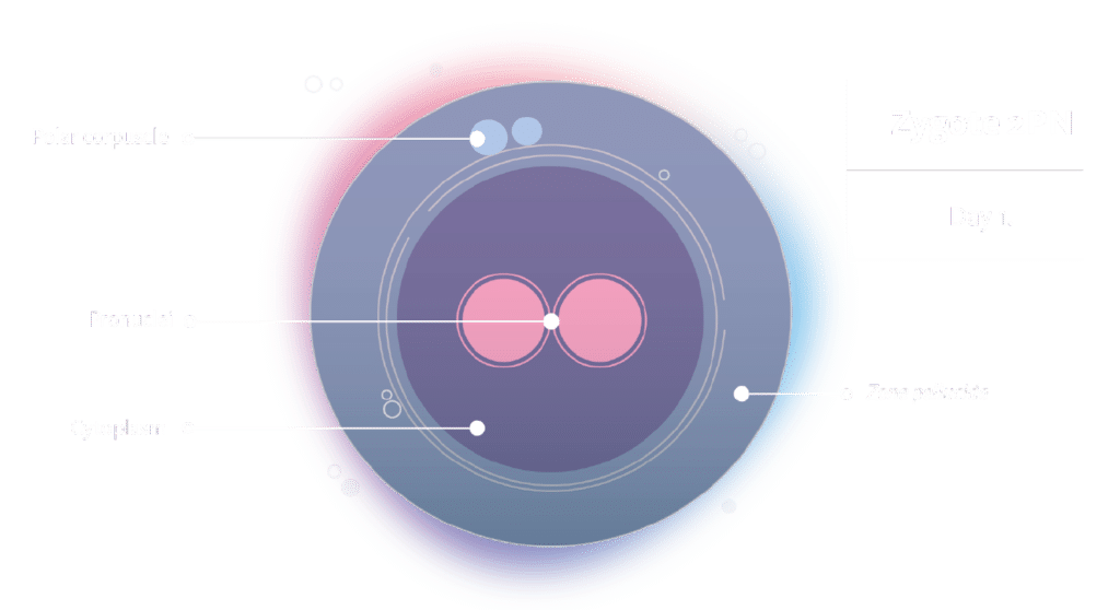
In assisted reproduction treatments, an oocyte is considered successfully fertilised when, 16-18 hours after in vitro fertilisation or microinjection, the zygote exhibits two pronuclei (2PN) and two polar bodies. Each pronucleus contains one set of chromosomes, with 23 chromosomes from the oocyte and 23 from the sperm. A few hours after their appearance, the pronuclei fuse and disappear, giving rise to the zygote nucleus with 46 chromosomes (23 pairs). These normal zygotes are cultured further to monitor embryo quality and development. In contrast, 5% of zygotes show one or more than two PN. These are considered abnormal and are discarded due to the elevated risk of abnormal ploidy (haploidy, triploidy, etc.).
How are the abnormal zygotes formed?
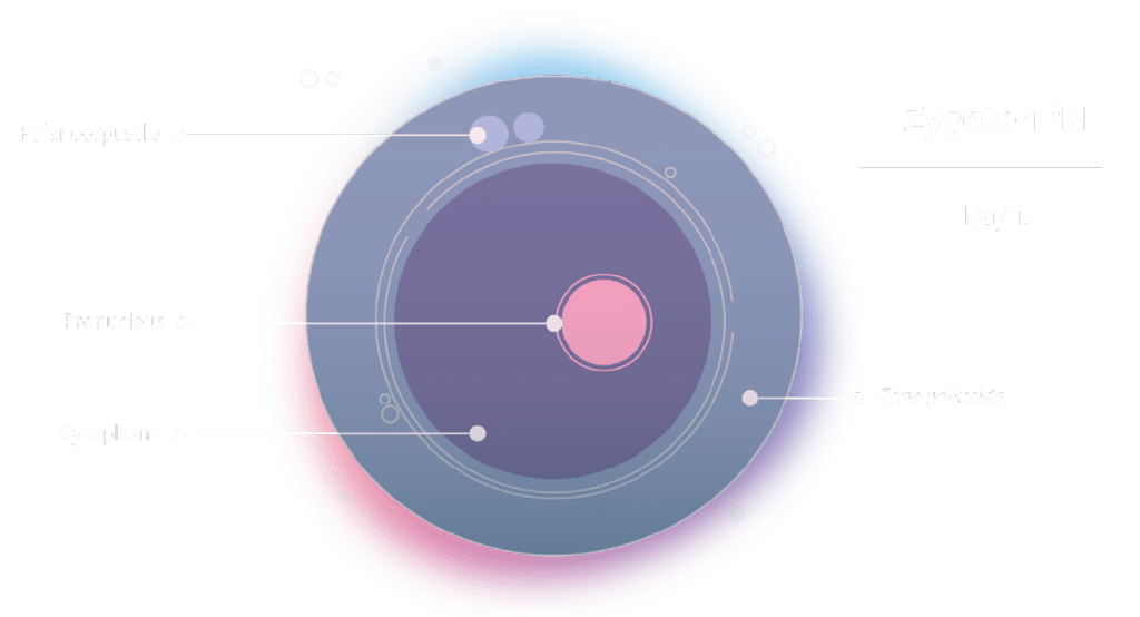
1PN zygotes can arise from parthenogenesis, activation of the nucleus of either the oocyte or sperm alone, complete extrusion of the female genetic material into a third polar body, abnormal nuclear envelope formation where the two genomes merge into a single pronucleus, or the organization of a nuclear envelope around only one of the parental genomes.
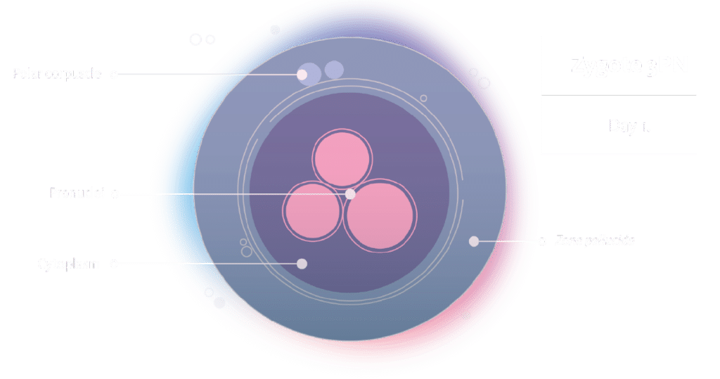
3 PN zygotes may occur due to fertilisation of the ovum by two spermatozoa, fertilization of a diploid sperm cell, failure to extrude the second polar body and the fragmentation or division of the first, or cleavage of one of the pronuclei.
Morphokinetics of abnormal zygotes
Frequently, these abnormal zygotes can undergo division and give rise to an apparently normal embryo. Nevertheless, both the literature and our data suggest that, in both scenarios, their developmental capacity is inferior to that of 2PN zygotes, with 1PN zygotes achieving the blastocyst stage at a lower rate.
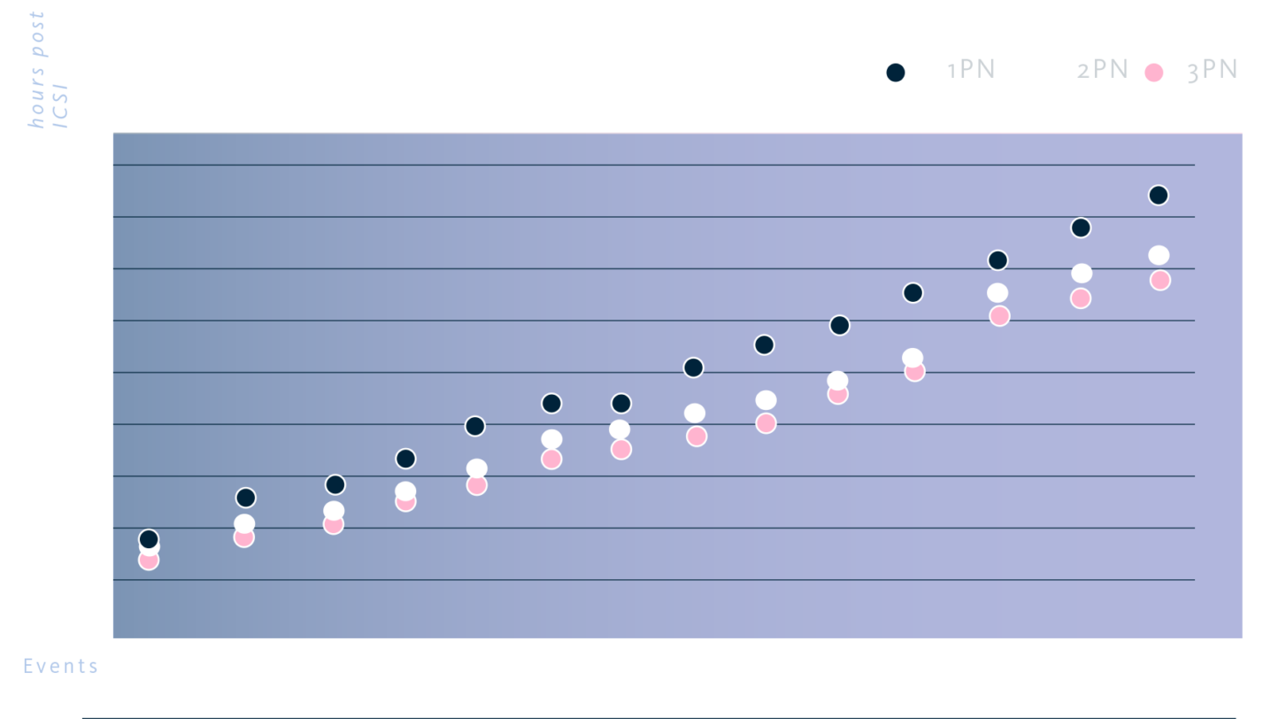
If the morphokinetic data, i.e. embryo division times and events, of 1PN, 2PN and 3PN zygotes are analysed and compared, it is observed that 3PN and 2PN embryos exhibit quite similar morphokinetics. However, 1PN zygotes show a slower pace, taking a longer time to reach any stage of embryo division.
Chromosomal composition of abnormal zygotes and gestation
1PN zygotes resulting from parthenogenetic activation of the oocyte or exclusive activation of the nucleus of one of the gametes will be haploid, indicating that they will contain only one set of chromosomes (n) from one of the parents. If a haploid embryo is transferred, it is likely failed to implant.
On the other hand, 3PN zygotes can be triploid, i.e. contain three sets of chromosomes. Although transferring them could lead to pregnancy, there is a high probability of miscarriage or the development of a molar pregnancy, posing a risk of choriocarcinoma.
1PN zygotes resulting from abnormal nuclear envelope formation, as well as 3PN zygotes originating from the splitting of one of the pronuclei, could potentially be diploid and have a normal chromosomal constitution. Consequently, they might be considered for reproductive purposes in cases where embryos from normal zygotes are not available.
Preimplantation genetic diagnosis of aneuploidy (PGT-A) allows us to ascertain the chromosomal endowment of embryos, but not their ploidy. In other words, an embryo can be euploid (with 46 chromosomes) but simultaneously diploid (2n), haploid (1n), or triploid (3n). Recently, several studies have been published wherein the use of single nucleotide polymorphisms (SNPs) enables the determination of embryo ploidy, as well as whether the chromosomes originate from the paternal or maternal side.
Thus, in a study presented at the ESHRE 2023 congress by Girardi et al, after PGT-A and SNP analysis of 318 embryos from abnormal zygotes, 42% were euploid. Among these, 36% of embryos from zygotes with 1PN were diploid, and 33% of those from zygotes with 3 or more pronuclei were diploid. In an earlier study by Capalbo et al, though fewer embryos were analysed, successful gestations were achieved by transferring euploid diploid embryos from abnormal zygotes.
These findings provide us with tools to harness abnormal zygotes and offer patients, who do not have embryos from normal zygotes more, opportunities to achieve a successful gestation.
Conclusions
Zygotes with 1 and 3 pronuclei can develop into embryos, although in a lower proportion than zygotes with 2. PGT-A, in conjunction with single nucleotide polymorphism (SNP) analysis, can effectively distinguish between euploid and non-euploid, diploid and non-diploid embryos, as well as identify the chromosomal endowment of maternal and paternal origin. Euploid embryos derived from abnormal zygotes have the potential to result in healthy gestations. Rescuing these embryos from cycles with a low number of embryos can increase the chances of gestation for these patients.
References
- Bradley, CK, Traversa, MV, Hobson, N, Gee, AJ, McArthur, SJ. 2017. Clinical use of monopronucleated zygotes following blastocyst culture and preimplantation genetic screening, including verification of biparental chromosome inheritance. Reprod. Biomed. Online 34, 567–574.
- Capalbo, A., Treff, N., Cimadomo, D., Tao, X., Ferrero, S., Vaiarelli, A., Colamaria, S., Maggiulli, R., Orlando, G., Scarica, C., Scott, R., Ubaldi, F. M., & Rienzi, L. 2017. Abnormally fertilized oocytes can result in healthy live births: improved genetic technologies for preimplantation genetic testing can be used to rescue viable embryos in in vitro fertilization cycles. Fertil Steril. 108(6):1007-1015.e3. doi:10.1016/j.fertnstert.2017.08.004
- Girardi L, Cogo F, Zambon P, Pergher I, Castellón JA , Patassini C, Rubio C. 2023. The incidence of different ploidy alterations in abnormally fertilized oocytes (AFO)-derived embryos. ESHRE Annual Meeting 2023. Human Reprod Volume 38, Supp 1
- Kai, Y, Moriwaki, H, Yumoto, K, Iwata, K, Mio, Y. 2018. Assessment of developmental potential of human single pronucleated zygotes derived from conventional in vitro fertilization. J. Assist. Reprod. Gen. 35, 1377–1384.
- Kemper JM, Liu Y, Afnan M, Mol BWJ, Morbeck DE. 2023. What happens to abnormally fertilized embryos? A scoping review. Reprod Biomed Online. 46(5):802-807. doi:10.1016/j.rbmo.2023.02.005
- Miravet-Valenciano J, Jimenez-Almazan J, Girardi L, Rodrigo L, Sato Y, Kayali R, Rubio C, Blesa D. 2023. Incidence of triploidy by NGS in trophectoderm biopsies from normally fertilized oocytes. 20th International Conference on Preimplantation Genetics. April 17-19, 2023, Paris.